





 Seraphina Hughes
 A new generation of molecular “GPS” technology is revolutionising our understanding of how cell receptors-particularly G protein-coupled receptors (GPCRs)-move, change shape, and transmit signals. By tracking these protein motions with unprecedented precision, scientists are gaining crucial insights into the workings of the body’s most important molecular switches, with direct implications for drug discovery and the treatment of diseases.
Introduction
Cell receptors are the gatekeepers of the body’s communication networks, translating external signals-such as taste, pain, hormones, and stress-into cellular responses. Among these, G protein-coupled receptors (GPCRs) stand out as the most versatile and widely targeted class, involved in nearly every physiological process and the action of about one-third of all approved drugs.
Despite their importance, the dynamic motions and conformational changes that underpin GPCR function have long eluded scientists. Traditional structural biology methods, such as X-ray crystallography, offer only static snapshots of these proteins, missing the critical transitions that occur as receptors activate, transmit signals, and reset. Now, by adapting principles similar to satellite GPS navigation, researchers have developed a method to track the motions of these proteins in real time, offering a dynamic view of their function at atomic resolution.
The New GPS for Proteins: Nuclear Magnetic Resonance (NMR) Tracking
How It Works
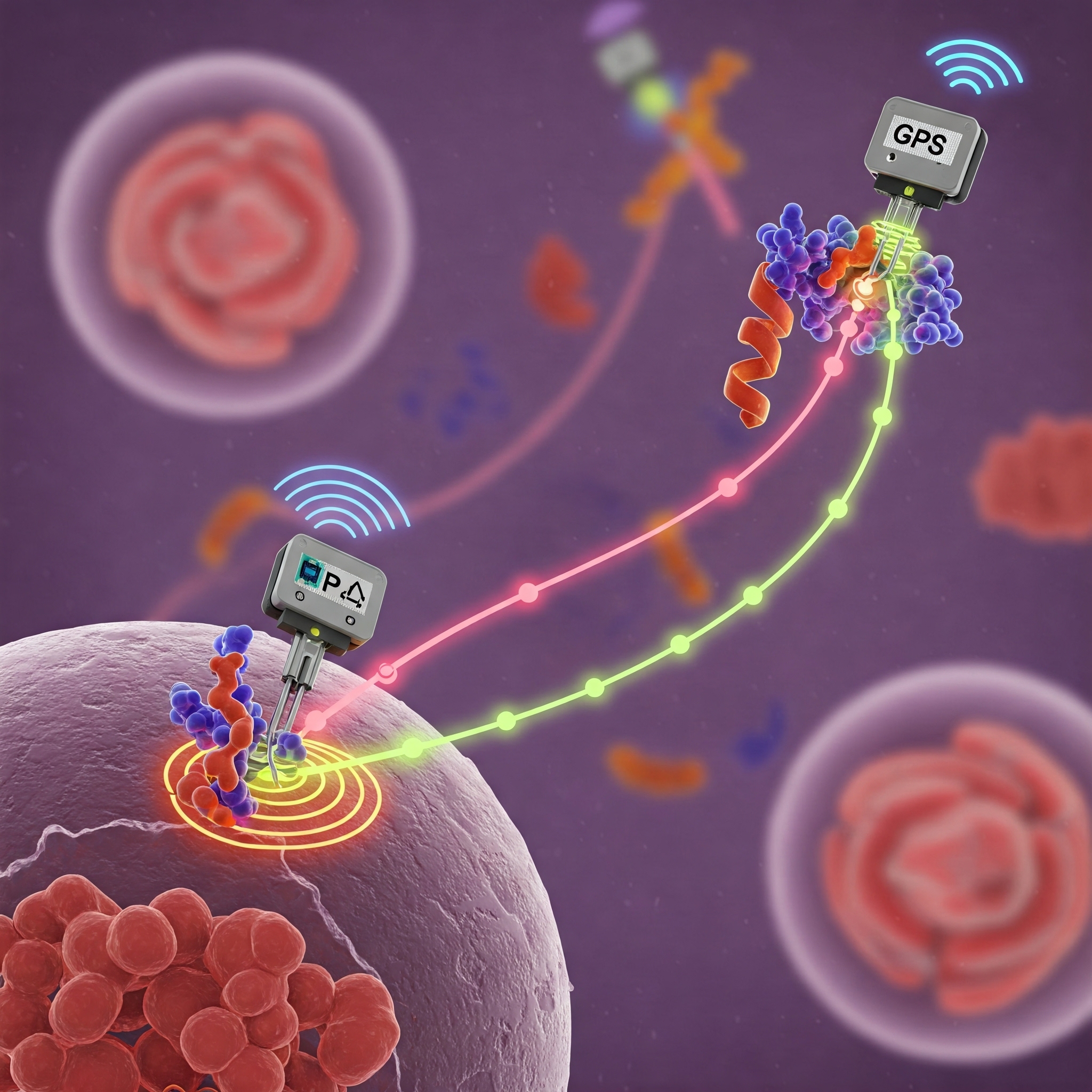
Researchers at the University of Basel have pioneered a technique that uses nuclear magnetic resonance (NMR) spectroscopy to monitor the atomic-scale movements of GPCRs. The approach involves attaching microscopic paramagnetic tags-acting as “GPS beacons”-to specific sites on the receptor protein. When placed in a magnetic field, these tags alter the local magnetic environment, producing signals that can be detected and precisely mapped by NMR.
By analysing how these signals change as the receptor interacts with different ligands (such as hormones or drugs), scientists can reconstruct the movements and conformational shifts of the protein in real time. This is analogous to how GPS satellites triangulate the position of a receiver on Earth, but at a molecular scale.
Advantages Over Traditional Methods
- Dynamic, Not Static: Unlike crystallography, which freezes proteins in a single state, NMR tracking captures the full range of motions and intermediate conformations.
- Atomic Precision: The method allows researchers to observe changes at the level of individual atoms, revealing subtle shifts that are crucial for function.
- Functional Context: By monitoring receptors in environments that mimic the cell membrane, the technique preserves the natural flexibility and complexity of the protein.
What GPCR Motions Reveal About Cell Signalling
The Architecture of GPCRs
GPCRs are integral membrane proteins with a characteristic seven-transmembrane helix structure. They respond to a wide variety of external signals, from neurotransmitters and hormones to sensory stimuli, by undergoing conformational changes that activate intracellular G proteins and trigger signalling cascades.
When a ligand binds to the extracellular side of a GPCR, it induces a series of structural rearrangements-most notably, an outward movement of certain transmembrane helices on the cytoplasmic side. This creates a binding cavity for the G protein, initiating the signal transduction process.
Tracking Activation and Signal Transmission
The new GPS NMR method allows scientists to observe these conformational changes as they happen. For example, researchers can see how different ligands stabilise distinct receptor states, how the receptor toggles between active and inactive forms, and how mutations or drugs alter its behaviour.
This level of detail is critical for understanding:
- Ligand Specificity: Why some drugs activate a receptor while others block it.
- Signal Bias: How different ligands can preferentially trigger certain signalling pathways, leading to specific physiological effects.
- Receptor Regulation: How receptors are desensitised, internalised, or recycled after activation.
Quantifying Receptor Motions: Mathematical and Statistical Models
Describing Motion Types
Advanced imaging and tracking methods, including fluorescence and NMR, reveal that receptor motions can be classified as:
- Brownian (Random) Motion: Typical of freely diffusing receptors in the membrane.
- Confined Motion: Movement restricted to specific membrane domains.
- Directed Motion: Active transport along cytoskeletal tracks or during endocytosis.
Statistical analysis of receptor trajectories often involves calculating the mean squared displacement (MSD):MSD(τ)=⟨[x(t+τ)−x(t)]2⟩MSD(τ)=⟨[x(t+τ)−x(t)]2⟩
where x(t)x(t) is the position at time tt and ττ is the time interval. The shape of the MSD curve helps distinguish between random, confined, and directed motion.
Classifying Dynamic States
Recent studies employ statistical hypothesis testing and machine learning to classify receptor motion into distinct categories based on trajectory data. For each segment of a receptor’s path, metrics such as maximal distance from the origin and motion standard deviation are computed and compared to reference distributions for Brownian or directed motion.
This classification not only reveals the diversity of receptor behaviours but also links specific motion patterns to functional states-such as activation, dimerisation, or interaction with signalling partners.
Implications for Drug Discovery and Medicine
Why GPCR Dynamics Matter
Understanding the real-time motions of GPCRs is crucial for drug development. Many drugs work by stabilising specific receptor conformations, but without knowledge of the full dynamic landscape, it is difficult to predict or optimise drug effects. The new GPS NMR technology provides a roadmap for designing molecules that precisely modulate receptor activity, potentially reducing side effects and improving efficacy.
For example, drugs like beta-blockers, painkillers, and diabetes treatments (such as semaglutide) all target GPCRs. Subtle differences in how these drugs interact with the receptor-and how they influence its motion-can lead to vastly different therapeutic outcomes.
Towards Personalised Medicine
By mapping the conformational preferences of GPCRs in different tissues, or in the presence of disease-associated mutations, researchers can tailor therapies to individual patients. This approach opens the door to more personalised and effective treatments for conditions ranging from heart disease to neurological disorders.
Broader Impact: Illuminating the Molecular Machinery of Life
Beyond GPCRs
While the current focus is on GPCRs, the GPS NMR method and related tracking technologies are being extended to other membrane proteins and molecular machines. This includes ion channels, transporters, and enzymes-all of which rely on dynamic motions for their function.
Integrating with Other Technologies
Combining NMR tracking with high-resolution fluorescence imaging, cryo-electron microscopy, and computational modelling provides a comprehensive toolkit for studying protein dynamics in situ. These multidisciplinary approaches are poised to transform our understanding of cellular signalling, disease mechanisms, and the development of next-generation therapeutics.
Summary
The advent of GPS-like tracking for proteins marks a turning point in cell biology and pharmacology. By illuminating the real-time motions of GPCRs and other receptors, scientists are uncovering the fundamental mechanisms that drive cellular communication and response. This dynamic view, made possible by innovations in NMR spectroscopy and statistical modelling, is not only solving long-standing mysteries in molecular biology but also charting a path towards smarter, safer, and more effective medicines. As the technology matures, the ability to “track” proteins in action promises to unlock new frontiers in our understanding of life at its most fundamental level.